Regular eye exams become more important as you reach your 40s and 50s. Not only do you need to keep pace with the changes in your vision by updating your prescription for glasses or contact lenses, but you also want to be certain there’s no vision problem beginning to  develop. Many of these problems have no physical symptoms until they’re quite advanced. You should have eye exams every year once you’ve reached 40 – and these exams are even more important if you have high blood pressure, diabetes or a family history of eye disease.
What Can You Expect at Your Eye Exam?
Each eye doctor has a routine, but most eye exams follow a similar pattern. First, your doctor will review your personal and family health history as you may be at special risk for eye problems if you have a family history of eye disease, diabetes, high blood pressure, or poor vision.
Then, your doctor will conduct tests to check for:
-
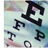
Vision - The doctor can check for shortsightedness (myopia), longsightedness(hyperopia), astigmatism and presbyopia. While you look at an eye chart, the doctor will measure your vision precisely, and, if necessary, determine a prescription for corrective lenses.
-
Coordination of eye muscles - The doctor will move a light in a set pattern to test your ability to see sharply and clearly at near and far distances, and to use both eyes together.
-
Side (peripheral) vision - The doctor will move an object at the edge of your field of vision to make sure you can see it.
-
Pupil response to light - The doctor will shine a light in your eye and watch the pupil's reaction.
-
Colour testing - The doctor will ask you to describe figures in a series of illustrations made up of numerous coloured dots or circles. This tests your ability to differentiate colours.
-
Eyelid health and function - The doctor will examine your eyelid, inside and out.
-
The interior and back of the eye - After dilating your eyes (by both using a few eye drops and dimming the lights so the pupils will widen), the doctor will use a special instrument called an ophthalmoscope to see through to the retina and optic nerve at the back of the eye. This is where clues to many eye diseases first show up.
-
Measurement of fluid pressure - The doctor will release a puff of air onto your eye using an instrument called a tonometer. This tests the pressure inside the eye, an early indicator of Glaucoma and other diseases.
What Can I Do to Self-Monitor My Vision Between Professional Visits?
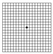
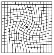
One way to self-monitor between professional visits is by looking at an Amsler grid. This is a pattern that resembles a checkerboard with a dot in the centre. While staring at the dot, you may notice that the straight lines in the pattern appear wavy. Or you may notice that some of the lines are missing. Print and take the quick vision test using the Amsler grid(72.3 KB, PDF). By looking at an Amsler grid regularly you can monitor any sudden changes in your vision. If you do notice any changes, contact your eye doctor right away.
|
Amsler grid - normal vision

|
Amsler grid - with AMD

|
Here's how:
-
Do the test with each eye separately by covering the eye that’s not being tested.
-
Hold the test grid directly in front of you, about 14 inches from your face, and look at the dot in the centre of the grid, not at the lines.
-
While looking at the dot, all the lines, both vertical and horizontal, should appear straight and unbroken.
-
If any of the straight lines appear wavy, or you notice that some of the lines seem to be missing, note their location on the grid for future reference.
Remember, this test is not meant to replace your regularly scheduled eye examinations. The best way to detect and monitor for conditions affecting the macula is for your eye care professional to use special instruments to examine the back of the eye.